4407 Manchester Ave # 204, Encinitas, CA 92024, United States
Upper Cervical Chiropractor in Encinitas
How It Works
The anatomy, the technique, and why precision matters.
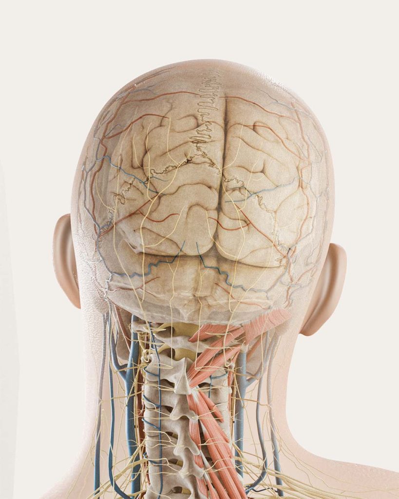
The upper cervical complex is the region at the base of the skull where the occiput, the atlas (C1), and the axis (C2) meet, along with the joints, ligaments, muscles, nerves, and blood vessels that pass through and around them. It is the most mobile region in the spine, and one of the most neurologically important.
What happens here can affect the entire body. The brainstem, the vagus nerve, the vertebral arteries supplying blood to the brain, and the sensory pathways for balance and head position all converge in this small region. The suboccipital muscles at the base of the skull are densely packed with sensory receptors and send constant feedback to the brain about head position and posture. Disrupt the alignment here and the downstream effects can reach a long way: tension and pain patterns, headaches, dizziness, balance and proprioceptive changes, jaw issues, postural compensations down the spine.
Nearly every neural pathway that the brain and body use to communicate passes through or near the atlas.
The atlas and axis are also the most freely moveable vertebrae in the spine, which makes them among the most easily disrupted. A fall, a car accident, a sports impact, or years of postural stress can shift their alignment. The root can be at the top of the neck even when the symptom lives somewhere else.
This is sometimes called an atlas subluxation. We use “misalignment” or “joint dysfunction” because the language is more descriptive.
The Blair technique
The Blair technique was developed by Dr. William G. Blair in Lubbock, Texas, starting in 1949. He noticed something that became the foundation of the method: the upper cervical joints are shaped differently in every person, and they are not symmetric from side to side within the same person. He built a system that accounts for that individual anatomy.
Three things define it:
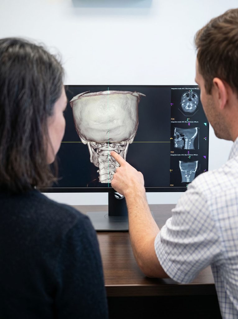
- Imaging-guided. Joint angles are mapped before any correction is made. In a modern clinic, with 3D CBCT.
- Custom to your anatomy. The angle, vector, and contact point are calculated from your imaging. No two adjustments are alike.
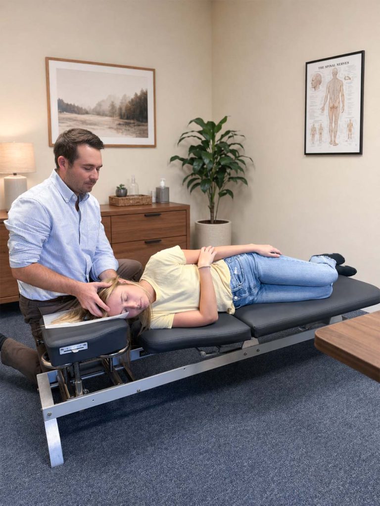
- Gentle and low-force. No twisting, cracking, or rotational manipulation.
Many patients are surprised by how light the adjustment feels. The amount of force used has nothing to do with whether the correction was effective.
The goal is not to adjust over and over. It is to restore alignment so the correction holds, the muscles can come out of guard mode, and the body can do what it is built to do.
Why precision matters
No two upper cervical spines are shaped alike, even from side to side within the same person. A generic adjustment vector that works for one patient will not fit the next. Without imaging, the adjustment is a guess. With it, the correction is specific: this contact, this angle, this direction, for this patient.
CBCT matters here because a 2D x-ray flattens the asymmetry the technique depends on. CBCT produces a true 3D model of the joints, with comparable or lower radiation than a standard cervical x-ray series.
This is also where the approach differs most from general chiropractic. General chiropractic typically addresses the full spine, with similar adjustments each visit, often based on feel. Upper cervical care is narrower and more specific: the focus stays on the upper neck, the decisions are guided by imaging and objective testing, and an adjustment happens only when the testing indicates it is needed.
Frequently Asked Questions
Is the Blair adjustment safe?
Yes. The technique avoids the high-force, rotational manipulations some patients are wary of. The adjustment is light, directional, and based on imaging of your individual anatomy.
Does it crack or pop?
No. There is no twisting, cracking, or rotational manipulation. Many patients are surprised at how light the correction feels.
How is this different from NUCCA or Atlas Orthogonal?
All three are upper cervical methods and share the same core principle: imaging guides the correction. They differ in how the imaging is taken and how the adjustment is delivered. NUCCA and Blair use hand-contact adjustments with the patient on their side. Atlas Orthogonal uses a percussion instrument. Blair stands out for highly individualized imaging-based adjustments tailored to each patient’s joint angles.
Will I need a lot of adjustments?
The aim is the opposite. The technique is designed so each correction has the best chance of holding. Many patients move from frequent visits early on to less frequent check-ins as the region stabilizes.
Does CBCT use a lot of radiation?
No. A single CBCT scan of the upper cervical spine is comparable to or lower than a standard cervical x-ray series, and significantly less than a medical CT scan.
Can children be adjusted with the Blair technique?
Yes. The technique is gentle enough to be appropriate for children, with adjustments made based on individual anatomy. As with adults, an adjustment is delivered only when testing indicates it is needed.
What conditions can the Blair technique help with?
The Blair technique addresses upper cervical misalignment, not specific conditions. That said, upper cervical misalignment can contribute to neck pain, headaches and migraines, dizziness, post-concussion symptoms, jaw tension, and balance issues. Whether the upper neck is contributing in your case is what the initial exam is for.